Skip to content
B. CADOT Research Website
Focus
Bibliography
Outreach
People
Scripts
SkyPad
Tools
Fancy
Personal Pics
Press
Where we are
Contact
Fancy
One PW1 interstitial cells (PICS) is followed over time and put in differentiation medium.
Cells transfected with RFP-utrophin and imaged with a 60x objective using a widefiled fluorescent microscope.
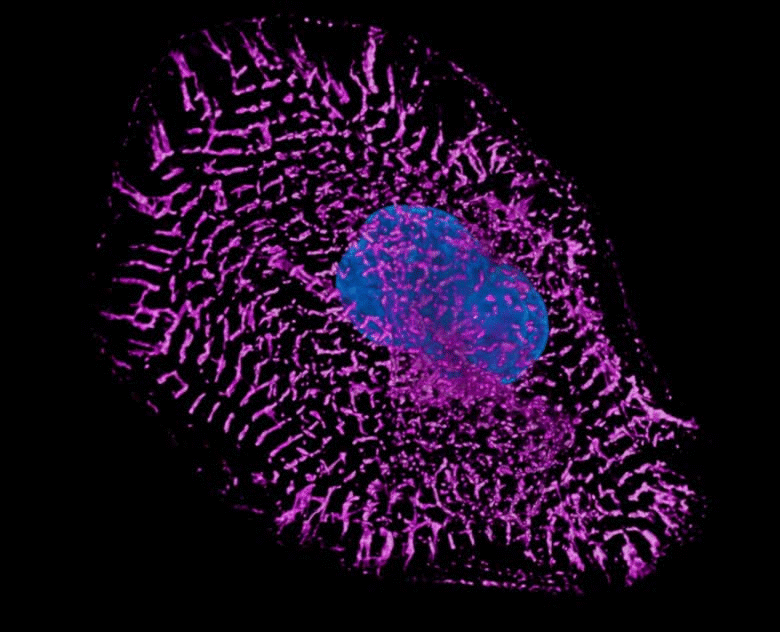
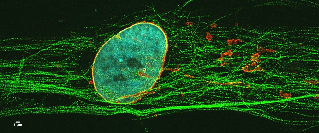
Emerin, Golgi and nucleus in a non-differentiated muscle cell
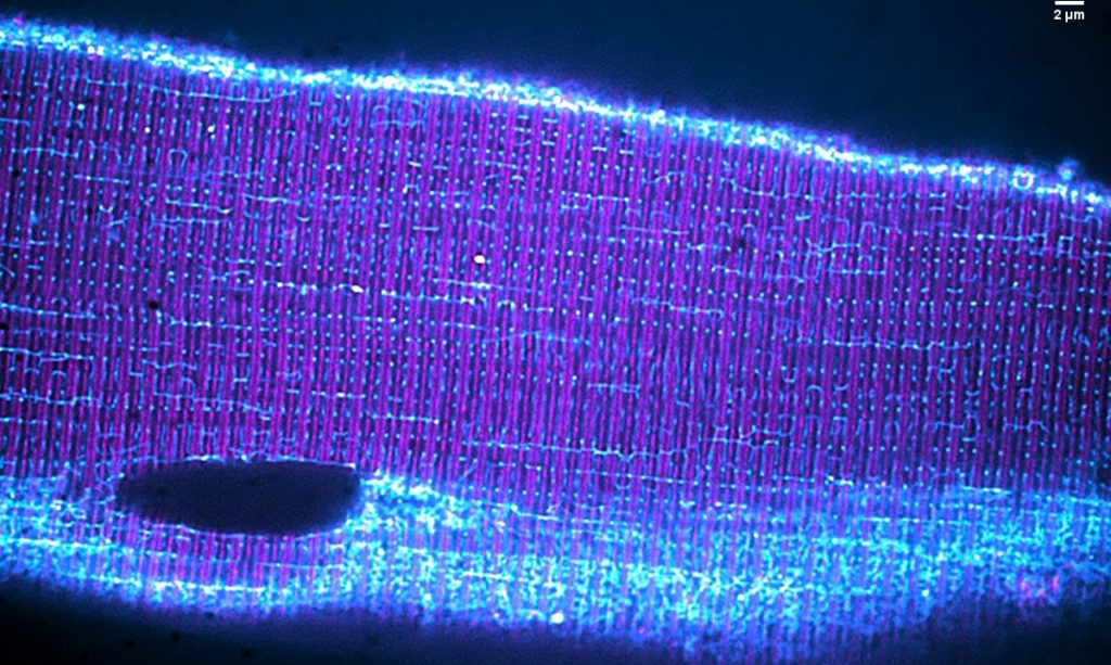
Microtubule gliding
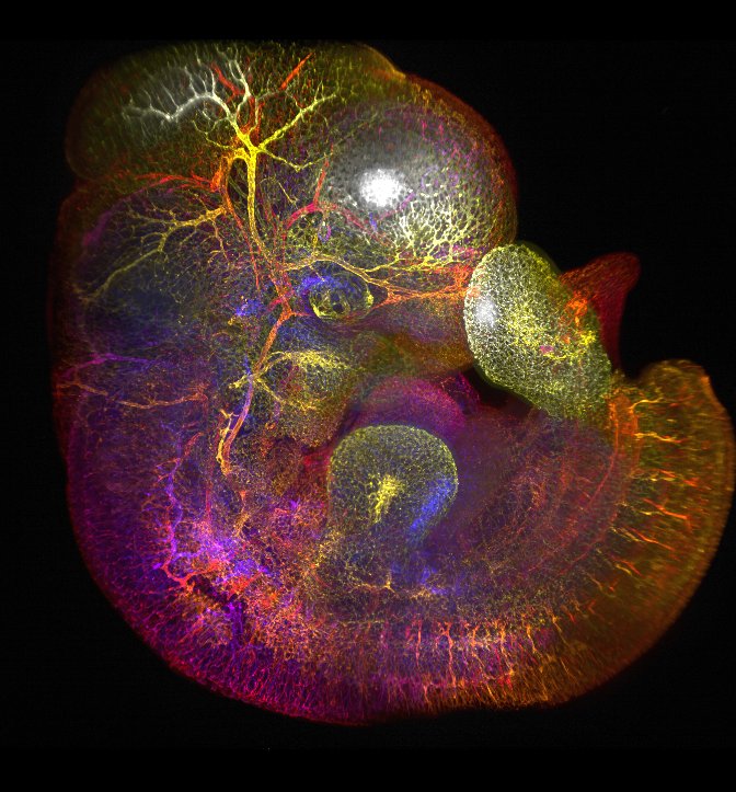
A PW1 interstitial cell (GFP) fuse with a satellite cell
C2C12 with GFP-expressing nuclei during the process of differentiation into myotubes
.
Partager :
Share on X (Opens in new window)
X
Share on Facebook (Opens in new window)
Facebook
Like
Loading…
Loading Comments...
Write a Comment...
Email (Required)
Name (Required)
Website
B. CADOT Research Website
Sign up
Log in
Copy shortlink
Report this content
Manage subscriptions
%d